-
The University
- Welcome
- Who we are
- Media & PR
-
Studying
- General
- Degree programs
- Campus life
-
Research
- Profile
- Infrastructure
- Cooperations
- Services
-
Career
- Med Uni Graz as an Employer
- Educational Opportunities
- Work Environment
- Job openings
-
Diagnostics
- Patients
- Referring physicians
- Health Topics
- Health Infrastructure

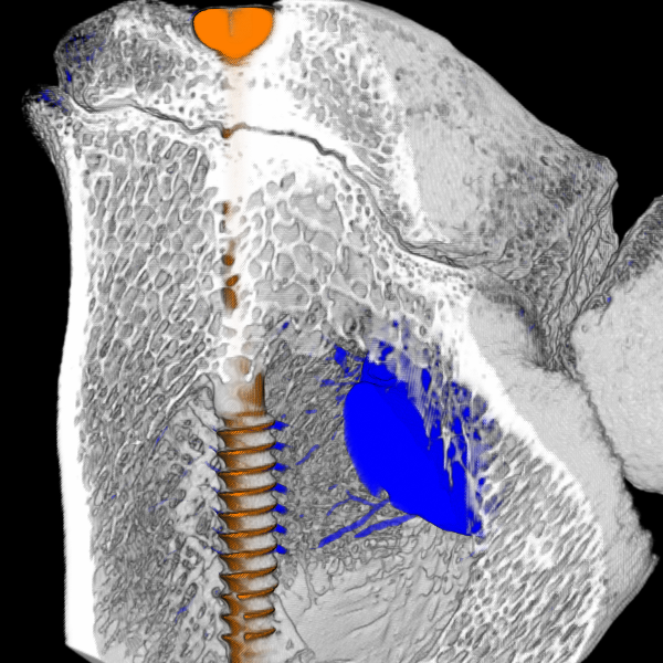
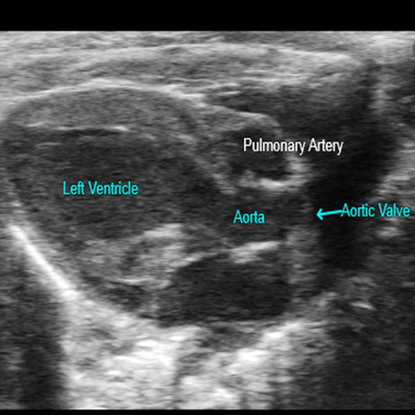
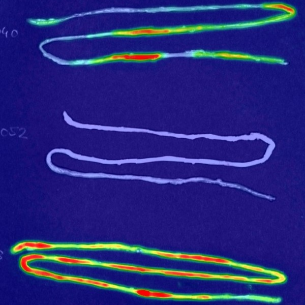
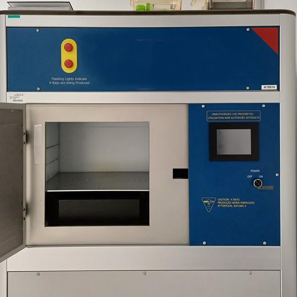
The Core Facility Preclinical Imaging (PCI) offers a state-of-the-art infrastructure and expertise for preclinical imaging addressing in vivo, ex vivo, and in vitro questions in basic and translational research. By applying a multimodal approach, continuous training, and scientific collaborations, research questions are examined from various perspectives while considering the 3R principles. The Core Facility offers preclinical imaging by MicroCT, MicroUltrasound und Optical Imaging as well as the use of a Biological Irradiator.
Our interdisciplinary team provides the know-how and experience required to obtain reliable and reproducible images and data. Researchers are supported continuously from project proposal submission through planning and execution to optimal data evaluation and storage.
The systems of the Core Facility PCI can be used individually or in combination to address a wide range of questions.
An initial meeting at the start of the project forms the basis for an optimal outcome. Within this framework, the precise research question, available analysis options, as well as the estimated time and cost requirements are coordinated. The establishment of the method, its execution, and data evaluation are carried out depending on the device either by the PCI core staff or by the project group itself.
Managing Director